Vascular Birthmarks
More than one in ten babies have red spots at birth or within the first few weeks of life.
Nevus flammeus
The most common type of vascular birthmark, these are located on the back of the neck most often, but can also appear on the forehead, eyelids, tip of nose, or upper lip. They are faint pink. The lesions on the face often resolve in the first few years of life, but the lesions on the back of the neck ("stork bite") commonly remain throughout life.
These birthmarks are harmless and require no treatment.
Hemangioma
This lesion does not usually appear immediately at birth, but becomes visible within the first few weeks of life. Unlike other vascular birthmarks, hemangiomas usually grow very rapidly. Growth generally begins during the first six weeks of life and continues for about six months to one year. After the first year, most hemangiomas will stop growing. They then begin to turn gray-white and slowly shrink. Half of all hemangiomas (50%) are flat by age five. Nine out of ten (90%) are flat by age nine. Many will completely go away, but often, a faint mark is left.
A hemangioma located near an eye, nose or mouth, or over the genitals or rectum, can cause special problems. These hemangiomas should be watched closely by your dermatologist, who will decide if further treatment is necessary.
Occasionally, a hemangioma that is growing or shrinking rapidly can form an open sore or ulcer. These sores are often painful, and can become infected. See your dermatologist for care soon.
Treatment:
There are several different treatments for hemangiomas. Most of the time, your dermatologist will choose to watch the lesion.
Rapidly growing hemangiomas were once treated with corticosteroid medication, but more recently a blood pressure medication called propranolol has been shown to provide good results. These treatments may carry side effects to be discussed with your specialist.
Pulsed dye laser can be used to prevent growth of hemangiomas and to remove superficial hemangiomas. Hemangiomas with sores that will not heal can also be treated with lasers.
Port wine stain
The port-wine stain occurs in 3 in 1,000 infants. Port-wine stains are visible at birth. They are flat and pink or red. Port-wine stains are found most often on the face, neck, arms or legs.
Unlike hemangiomas, port-wine stains do not resolve by themselves, and are permanent. Later in life, port-wine stains may become thick and develop small blood vessel growths called vascular blebs.
Port-wine stains on the forehead, eyelids or both sides of the face, can be associated with glaucoma and/or seizures. Glaucoma is an increased pressure within the eye that left untreated, can cause blindness. These complications occur in less than half of those with port-wine stains of the forehead and eyelids. All infants with a port-wine stain in those areas should have a thorough eye examination, and if indicated, further brain imaging.
Treatment:
The preferred treatment is pulsed dye laser, which targets blood vessels. Many treatments may be needed, at 2-3 month intervals. Some port wine stains will respond very well, while others may hardly change. This can often be predicted before treatment is started.
Immediately after treatment, the area will appear bruised for 10 days or so. Makeup can be applied. Sun exposure must be avoided before and after treatment. Blisters are uncommon and should be reported to the doctor. In rare cases, laser treatment can cause temporary changes in skin color. In very rare cases, permanent color change or scars may occur. The treatment is mildly uncomfortable; a topical anesthetic cream like EMLA can be applied an hour before treatment.
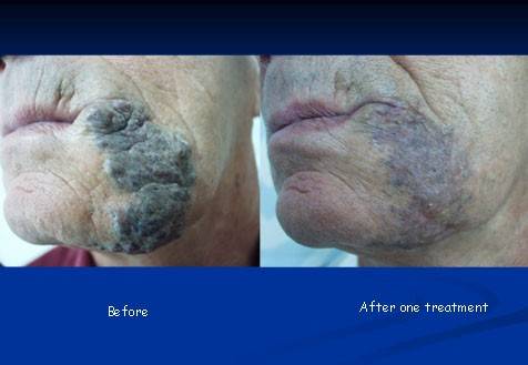
Venous malformations
These lesions are present at birth but may become evident mush later in life. They are typically blue-gray or purple in color and are deep and palpable. Treatment depends on the location and extent of the malformation and may involve several approaches, including laser, surgery, and injection of agents that close the blood vessels from within.
Adapted from the American Academy of Dermatology
The information above is a partial and general information and cannot replace the needed specific medical examination and consultation